How to Use Ultrasound Machine: Definition & Purposes
Ultrasound machine is a special medical device used in ultrasonography for diagnostic and therapeutic purposes. Ultrasonography, commonly known as ultrasound, is a popular medical imaging technique used in physical therapy and diagnostics, and which uses high-frequency sound pulses and their echoes to specific body regions. Ultrasonic waves are specifically helpful in visualizing or imaging subcutaneous or internal body structures including muscles, tendons, vessels, joints or internal organs for any pathology or lesions.
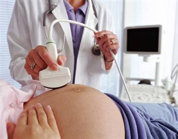
Ultrasound machine is constantly becoming very popular for its diagnostic and therapeutic qualities. It doesn’t matter whether you’re pregnant and your obstetrician would like to monitor a developing fetus or determine the due date, or you’re experiencing blood circulation problems in your limb or heart and your physician needs to check your blood flow, ultrasound is always at your service. Ultrasound machines are successfully helping patients in reducing pain and improving the healing process, such as generating heat deep into muscles or breaking up minor calcifications and bruises by creating localized vibrations, increasing nutrients & oxygen and reducing swelling.
How to Use Ultrasound Machine: What Does It Consist Of & How Does It Work?
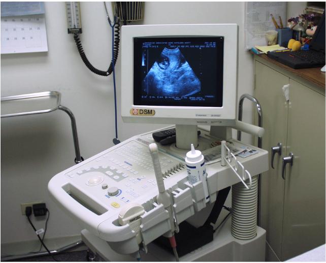
A basic ultrasound machine consists of the following parts:
- transducer probe (sends & receives the sound waves);
- transducer pulse controls (changes the pulses’ amplitude, frequency and duration);
- central processing unit (does the calculations);
- display (displays the images taken from the CPU-processed ultrasound data);
- disk storage device (stores the acquired images);
- keyboard & cursor (used to input data and take the measurements);
- printer (prints the image).
If you’re about to use an ultrasound machine, remember the following steps and procedures to be followed:
- remove clothes or bandages that may impede ultrasonic waves from penetrating into the afflicted tissue or organ;
- apply hypo-allergic gel & anti-inflammatory cream to the area to be treated;
- turn on the ultrasound unit (place the transducer probe on the gel and work it in circular motion for 5-10 minutes):
- the machine uses a transducer probe for transmitting high-frequency sound waves into the patient’s body;
- sound pulses travel into the patient’s body and hit a boundary between tissues;
- some sound waves will get reflected from the boundary back to the probe, while others continue to travel until another boundary is reached and then they get reflected as well;
- the transducer picks up the reflected sound pulses and relays them to the machine;
- the ultrasound machine uses the sound speed in tissue and each echo’s return time to calculate the distance from the probe to the organ or tissue;
- the ultrasound unit forms a two-dimensional image, which is a display of the distances and intensities of the echoes;
- turn the ultrasound off & wipe the hypo-allergenic gel away.

